Sciences du vivant
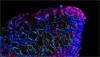
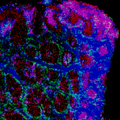
La spectroscopie Raman est employée avec succès dans l’analyse des cellules et tissus humains, animaux et végétaux.
- Biotransformation
- Analyse structurelle de protéines/peptides
- Microbiologie
- Administration de médicaments in vitro et in vivo
- Recherche sur le cancer / pathologie
- Biologie redox
- Médecine régénérative
- Maladies neurodégénérescentes et associées au vieillissement
- Recherche sur les biocarburants et l’agriculture
- Lipidologie
- Métabolomique
- Biologie du développement
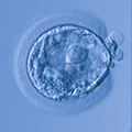
- Biologie de la reproduction
- Virologie
Cliquez sur les liens ci-dessous pour découvrir comment nous pouvons aider dans vos applications des sciences du vivant.
Nous sommes là quand vous en avez besoin
Plus d’infos sur ce domaine d’application ou sur une application non mentionnée ici, n’hésitez pas à nous contacter.
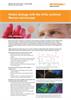
Contacter notre équipe ApplicationsWebinaire - La spectroscopie Raman de résonance dans la recherche biologique redox
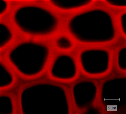
La spectroscopie Raman de résonance (RR) constitue l’outil idéal pour la recherche en biologie redox. Elle révèle non seulement une sensibilité élevée aux protéines héminiques, mais elle permet également de déterminer in situ leurs états d’oxydation et d’oxygénation (solution, organites, cellules et tissus). L’imagerie RR procure à la fois des informations chimiques et spatiales, permettant d’établir des corrélations entre la répartition des protéines héminiques, l’état d’oxydation et la fonction des protéines/cellules.
Regarder le webinaireTéléchargements : Sciences du vivant
-
 Brochure: Biological analysis using Raman spectroscopy and imaging [en]
The domain of biological research is shaped by our ability to peer into the world of the small. Simply seeing microscopic biological samples is useful, but by utilising Raman spectroscopy we can surpass sight into the molecular realm… and beyond! Download this brochure to discover the wealth of biological applications made possible by Renishaw's Raman systems.
-
 Notes d’applications : Biologie rédox avec le microscope Raman confocal inVia [it]
La spectroscopie Raman est sensible à la présence d’hémoprotéines et est idéale pour l’étude de la biologie rédox sans avoir à prévoir d’isolation ou de coloration. La rédox (oxydo-réduction) des hémoprotéines est étroitement liée aux fonctions des protéines (transport et stockage de l’oxygène, transport des électrons et inhibition des radicaux libres). En utilisant la spectroscopie Raman pour élucider les états de rédox au sein des systèmes biologiques, les chercheurs peuvent étudier la dynamique de rédox et ses effets sur la régulation de la santé et les maladies.
-
 Application note: Raman imaging for biological applications. No stains. No labels. [en]
Raman spectroscopy is an information-rich, label-free, non-invasive imaging technique that is ideal for life sciences research. It uses laser light scattering to provide a chemical fingerprint at each point of the analysed area and identifies the molecules present in samples.
-
 Product note: Microplate mapping with Renishaw Raman system's [en]
Renishaw’s microplate mapping package enables researchers to use Renishaw’s Raman spectroscopy products to rapidly and easily analyse material contained in microplates.

-
Raman chemical imaging for life sciences [en]
Short movie to demonstrate the benefits of using the inVia confocal Raman microscope for powerful, flexible, Raman Imaging for life sciences applications.